Arm Muscles Diagram Anterior : Forearm Muscles Anatomy Anatomy Drawing Diagram : This layer contains only one muscle, the flexor digitorum.
Arm Muscles Diagram Anterior : Forearm Muscles Anatomy Anatomy Drawing Diagram : This layer contains only one muscle, the flexor digitorum.. Arm anterior muscles labeled 3d illustration. Muscles of anterior (flexor) compartment of arm, their origin, insertion, action/s and nerve supply are as follows superior ulnar collateral branch of brachial artery. In general, these are the flexors of the wrist and fingers and pronate the forearm. Flexion of the forearm is achieved by a group of three additionally, the biceps brachii operates as a supinator of the forearm by rotating the radius and moving the palm of the hand anteriorly. Shoulder muscles chest muscles thigh muscles arm muscle anatomy muscle diagram bicep muscle yoga anatomy medical anatomy there are anterior muscles diagrams and posterior muscles diagrams. Produce wrist and/or finger flexion. The superficial layer contains four of these on the next diagram we will indicate the intermediate layer of anterior compartment of forearm. Pectoralis major, and anterior fibers of the deltoid. This layer contains only one muscle, the flexor digitorum. Thick triangular muscle drawing the arm away from the median axis of the body and directing it toward the front and back until it is horizontal. The anterior compartment of the arm is also known as the flexor compartment of the arm as its main action is that of flexion. Coracobrachialis brachialis biceps brachii coracobrachialis: Tutorials and quizzes on muscles that act on the arm/humerus (arm muscles: The arm muscles comprise five muscles, which mainly act to flex and extend the forearm. These muscles are all innervated by the musculocutaneous. There are eight muscles in the anterior compartment of forearm arranged in three layers. You can see it running just underneath the biceps and it inserts onto the humerus. This module is a comprehensive and affordable learning tool for medical students and residents and especially for rheumatologists, orthopedic surgeons and radiologists. Arm anatomy diagram for artists. Learn the muscles of the arm with free quizzes, diagrams and worksheets. Adbucts scapula and rotates it downward. In general, these are the flexors of the wrist and fingers and pronate the forearm. Introduction to functional anatomy of the arm muscles: The deltoid consists three sets of fibers: Usually as one muscle contracts (or shortens), the opposing muscle (known as the antagonist) elongates and vice versa. Arm anatomy diagram for artists. Arm anterior 3d illustration project. Tutorials and quizzes on muscles that act on the arm/humerus (arm muscles: Muscles that cross the elbow (moving the forearm) (anterior) 1) deltoid (visible, but not part of this group as it moves arm from the shoulder). Shoulder muscles chest muscles thigh muscles arm muscle anatomy muscle diagram bicep muscle yoga anatomy medical anatomy there are anterior muscles diagrams and posterior muscles diagrams. Draw labelled diagram showing branches of profunda brachi artery. The serratus anterior acts to pull the scapula forward around the thorax. This is the coracoid process. This module is a comprehensive and affordable learning tool for medical students and residents and especially for rheumatologists, orthopedic surgeons and radiologists. The serratus anterior is a muscle that originates on the surface of the 1st to 8th ribs at the side of the chest and inserts along the entire anterior length of the medial border of the scapula. The muscles of the upper arm are split into anterior and posterior compartments. The lower half of the front of the humerus including both the anteromedial and anterolateral surface and the anterior border insertion: Although the majority of the muscle mass is located anteriorly to the humerus, it has no attachment. The biceps brachii, the brachialis and the coracobrachialis. Arm anatomy diagram for artists. How to view the anatomical labels. The deltoid consists three sets of fibers: From anterior distal humerus d. In this image, you will find hand and forearm muscle anatomy, humerus, extensor carpi radialis longus muscle, anconeous muscle, lateral antebrachial cutaneous nerve, ulna, radial nerve, flexor carpi ulnaris, median nerve, brachioradialis muscle, medial antebrachial cutaneous nerve in it. Draw labelled diagram showing branches of profunda brachi artery. The serratus anterior is a muscle that originates on the surface of the 1st to 8th ribs at the side of the chest and inserts along the entire anterior length of the medial border of the scapula. Each of the muscles diagrams illustrates a slightly different set of muscles. Although the majority of the muscle mass is located anteriorly to the humerus, it has no attachment. Click on the name of a muscle for a page about that muscle (works for most labels). The lower half of the front of the humerus including both the anteromedial and anterolateral surface and the anterior border insertion: There are eight muscles in the anterior compartment of forearm arranged in three layers. The anterior compartment of the arm is also known as the flexor compartment of the arm as its main action is that of flexion. Learn the muscles of the arm with free quizzes, diagrams and worksheets. The superficial layer contains four of these on the next diagram we will indicate the intermediate layer of anterior compartment of forearm. See anterior view in : The muscles of the arm anatomical chart does an exemplary job of examining the individual muscles that make up this area of the human body, and included in the dozen or more beautiful illustrations are views of the dorsal area, thorax, triceps, biceps, brachialis, serratus anterior, promator and. Start studying arm anterior muscles. Draw labelled diagram showing branches of profunda brachi artery. Arm muscles with portions of arteries and nerves muscles of arm: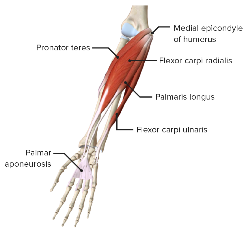
This module is a comprehensive and affordable learning tool for medical students and residents and especially for rheumatologists, orthopedic surgeons and radiologists.

The muscles labelled in the anterior muscles diagram shown above are listed in bold in the following table

Learn the muscles of the arm with free quizzes, diagrams and worksheets.
This layer contains only one muscle, the flexor digitorum arm muscles diagram. Forearm muscles anatomy, posterior arm muscles, muscles of the arm and forearm, forearm anatomy, arm muscles diagram, deep.
0 Comments:
Posting Komentar